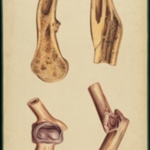
Teaching watercolor comparing the break of a human humerus at the sixth week of recovery to three broken dog tibias on the tenth and eighteenth days of recovery
Teaching watercolor comparing the break of a human humerus at the sixth week of recovery to three broken dog tibias on the tenth and eighteenth days of recovery
Description:
After Edward Stanley's Illustrations of the effects of disease and injury of the bones, plates 23 and 24 Large watercolor comparing the healing of fractures in humans and animals. The leftmost bone is a human humerus shown in cross section, with osseous tissue surrounding the break. The three right images are dog tibias, with the middle two at ten days and the far right at eighteen days after the break, in both cross section and exterior views. Watercolor is framed in green sewn textile, with metal grommets in each of the four corners.
The Harvard Medical Library does not hold copyright on all the materials in the collection. For use information, contact the Warren Anatomical Museum Curator at chm@hms.harvard.edu
Contact host institution for more information.
Notes:
Henry Jacob Bigelow employed artist Oscar Wallis exclusively from 1848 - 1854 to paint a series of large teaching watercolors to illustrate Bigelow's lectures at Harvard Medical School. Wallis painted the teaching diagrams from local subjects and from the atlases of established medical authorities. The effort cost Bigelow $6,000. In 1890 Bigelow presented the watercolors to Reginald H. Fitz to be used in the Harvard Medical School's Department of Anatomy. The watercolors were transferred into the Warren Anatomical Museum between 1890 and 1930.