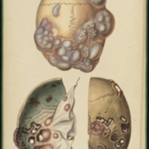
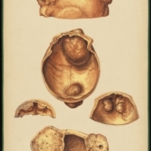
After Jean Cruveilhier's Anatomie pathologique du corps humain, vol. 2, liv. 33, pl. 4 Large watercolor showing two views of a skull covered in bone neoplasms. The top image shows the skull from the top, with the soft tissues of the tumor still attached, seen as bulbous masses of gray tissue. In the bottom left is a view of the interior of the skull, with holes and markings and changes in texture caused by the tumors. In the bottom right is the exterior of the skull with the soft tumors removed, showing the star-like injuries left in the bone. Watercolor is framed in green sewn textile, with metal grommets in each of the four corners.
The Harvard Medical Library does not hold copyright on all the materials in the collection. For use information, contact the Warren Anatomical Museum Curator at chm@hms.harvard.edu
Contact host institution for more information.
Notes:
Henry Jacob Bigelow employed artist Oscar Wallis exclusively from 1848 - 1854 to paint a series of large teaching watercolors to illustrate Bigelow's lectures at Harvard Medical School. Wallis painted the teaching diagrams from local subjects and from the atlases of established medical authorities. The effort cost Bigelow $6,000. In 1890 Bigelow presented the watercolors to Reginald H. Fitz to be used in the Harvard Medical School's Department of Anatomy. The watercolors were transferred into the Warren Anatomical Museum between 1890 and 1930.