Teaching watercolor of scar tissue, tumors, and abnormality of the nerves
Description:
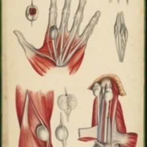
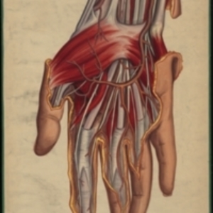
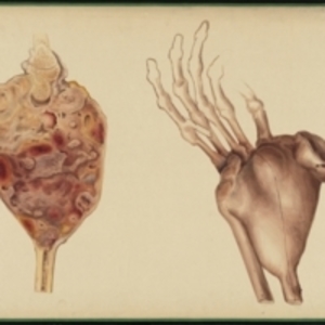
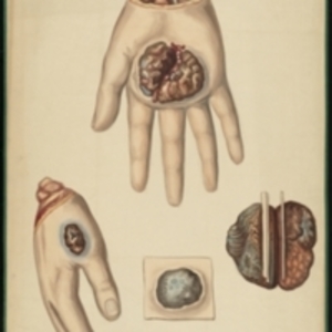
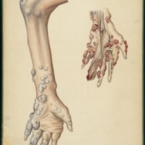
After Jean Cruveilhier's Anatomie pathologique du corps humain, vol. 2, liv. 35, pl. 2 Large watercolor showing several different nerves. In the upper left is a tumor on the nerve leading to the index finger of the hand. It is seen both dissected, and in its place on the hand, which shows only bones, muscles, and nerves. In the upper right are two views of an abnormal nerve fragment. In the lower left is another tumor on the nerve, both the exterior view in situ surrounded by muscle, exterior view isolated, and dissected view. On the lower right is scar tissue on a nerve on an amputated limb. Watercolor is framed in green sewn textile, with metal grommets in each of the four corners.
The Harvard Medical Library does not hold copyright on all the materials in the collection. For use information, contact the Warren Anatomical Museum Curator at chm@hms.harvard.edu
Contact host institution for more information.
Notes:
Henry Jacob Bigelow employed artist Oscar Wallis exclusively from 1848 - 1854 to paint a series of large teaching watercolors to illustrate Bigelow's lectures at Harvard Medical School. Wallis painted the teaching diagrams from local subjects and from the atlases of established medical authorities. The effort cost Bigelow $6,000. In 1890 Bigelow presented the watercolors to Reginald H. Fitz to be used in the Harvard Medical School's Department of Anatomy. The watercolors were transferred into the Warren Anatomical Museum between 1890 and 1930.