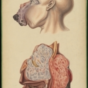
Teaching watercolor of exterior and interior views of a large bone tumor of the upper jaw which filled the cavities of the nose and orbits and extended into the cranium
Teaching watercolor of exterior and interior views of a large bone tumor of the upper jaw which filled the cavities of the nose and orbits and extended into the cranium
Description:
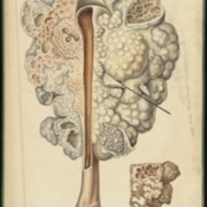
After Edward Stanley's Illustrations of the effects of disease and injury of the bones, plates 13 and 17 Large watercolor showing exterior and interior views of a large tumor afflicting a 14-year-old boy. The top image shows the head of the boy and the outside of the tumor, with the skin ulcerating and the eyes bulging. A large purple growth, with areas of red and black, extends from the nose down over the mouth. The cheeks and jaw are distorted. The lower image shows the interior of the tumor in cross section. The teeth and upper jaw are visible, and the tumor is divided into two sections - one that is grey with patches of yellow, and the other which is pink with a black exterior. Watercolor is framed in green sewn textile, with metal grommets in each of the four corners.
The Harvard Medical Library does not hold copyright on all the materials in the collection. For use information, contact the Warren Anatomical Museum Curator at chm@hms.harvard.edu
Contact host institution for more information.
Notes:
Henry Jacob Bigelow employed artist Oscar Wallis exclusively from 1848 - 1854 to paint a series of large teaching watercolors to illustrate Bigelow's lectures at Harvard Medical School. Wallis painted the teaching diagrams from local subjects and from the atlases of established medical authorities. The effort cost Bigelow $6,000. In 1890 Bigelow presented the watercolors to Reginald H. Fitz to be used in the Harvard Medical School's Department of Anatomy. The watercolors were transferred into the Warren Anatomical Museum between 1890 and 1930.