Teaching watercolor of a piece of tissue and a microscopic view
Description:
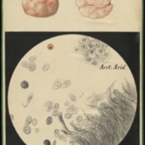
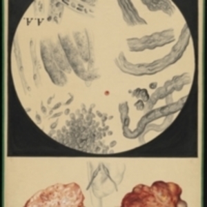
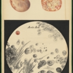
Possibly from local Boston patient Large watercolor showing a piece of tissue removed from a body. The tissue is round and pale in color, with red vessels on the surface. At the top of the painting is an exterior view and an interior view. The bottom part of the painting shows a round microscopic view, framed in black. In the view are round cells of various kinds, a red blood cell for scale, and long thin strands of tissue in the lower right. Watercolor is framed in green sewn textile, with metal grommets in each of the four corners.
The Harvard Medical Library does not hold copyright on all the materials in the collection. For use information, contact the Warren Anatomical Museum Curator at chm@hms.harvard.edu
Contact host institution for more information.
Notes:
Henry Jacob Bigelow employed artist Oscar Wallis exclusively from 1848 - 1854 to paint a series of large teaching watercolors to illustrate Bigelow's lectures at Harvard Medical School. Wallis painted the teaching diagrams from local subjects and from the atlases of established medical authorities. The effort cost Bigelow $6,000. In 1890 Bigelow presented the watercolors to Reginald H. Fitz to be used in the Harvard Medical School's Department of Anatomy. The watercolors were transferred into the Warren Anatomical Museum between 1890 and 1930.