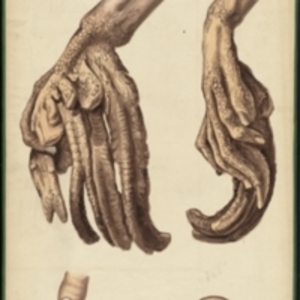
Teaching watercolor of a right hand with a skin disease that causes scaly skin and horn-like growths
Description:
After Jean Cruveilhier's Anatomie pathologique du corps humain, vol. 1, liv. 7, pl. 6 Large watercolor showing several views of a right hand affected by a skin disease. The surface of the skin is brown and scaly, and the hands are deformed. Skin and nail are extended into long, curved horns. At the bottom are two details of the tip of a finger, one from the exterior and one as a cross section showing the growth of the abnormal nail. Watercolor is framed in green sewn textile, with metal grommets in each of the four corners.
The Harvard Medical Library does not hold copyright on all the materials in the collection. For use information, contact the Warren Anatomical Museum Curator at chm@hms.harvard.edu
Contact host institution for more information.
Notes:
Henry Jacob Bigelow employed artist Oscar Wallis exclusively from 1848 - 1854 to paint a series of large teaching watercolors to illustrate Bigelow's lectures at Harvard Medical School. Wallis painted the teaching diagrams from local subjects and from the atlases of established medical authorities. The effort cost Bigelow $6,000. In 1890 Bigelow presented the watercolors to Reginald H. Fitz to be used in the Harvard Medical School's Department of Anatomy. The watercolors were transferred into the Warren Anatomical Museum between 1890 and 1930.